There is an important diagnostic tool that can make a significant difference in how we understand and treat TMJ disorders—the Panorex. This advanced imaging technique is a great screening tool that can help to identify issues that have to do with the TMJ (temporomandibular disorder) that may not be visible with traditional X-rays.
But many patients don’t want to have a panorex taken because of “radiation exposure”. So let me put this quickly into context:
A typical panorex = about 10 units of radiation (10 µSv)
Comparing this to daily everyday sources, here are some common ones:
Daily background radiation = 10 units
Chest x-ray = 100 units
1 serving of Brazil nuts = 5 units
Daily radiation from food = 1 unit
Sleeping next to someone = 10 units
So when we consider the fact that many people sleep next to someone, consume food, and have background exposure, all these amount to much more radiation on a daily basis than one panorex. Now here is what a panorex can help us see, and you will be surprised by some of what can be seen on a panorex that can greatly help up prevent, diagnose early, and manage TMJ issues before they become unmanageable and painful.
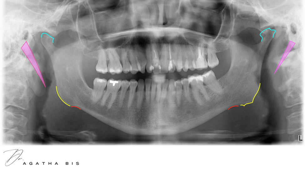
When looking at a panorex, most people think that dentists just see teeth. There may be impacted wisdom teeth, or decay, or bone loss. A panorex can help clinicians detect cancer, tumours, cysts, and much more. When it comes to TMJ Disorder, a panorex is invaluable. There are 4 key signs that can be seen on a panorex that are clear signs of parafunction and TMJ Disorder, before you have any kind of symptoms.
1. Notching
Notching at the angle of the mandible refers to an indentation or concavity that can be seen on a panorex, or sometimes palpated in the lower jaw. It looks like a concavity or ‘notch’ and forms an ante-gonial notch at the lower surface of the mandible. This is a sign of chronic clenching or grinding, which can lead to jaw pain and other TMD related symptoms.
2. Remodelling
Remodelling at the angle of the mandible refers to the process of bone reshaping or structural changes in the lower jaw area, typically again, as a result of chronic clenching or grinding. Typically, this looks like a thickening or extra bone at the angle of mandible and is a protective response to the masseter muscle contraction forces.
3. Condylar changes
Condylar changes in TMJ refer to alterations in the shape, size, or structure of the mandibular condyle, that result from degenerative joint disease, trauma, or inflammatory conditions. This could include flattening, osteophyte formation, erosions, cysts, and other degenerative changes.
4. Stylohyoid ligament calcifications
The styloid process is a slender, pointed projection of bone extending downward from the temporal bone of the skull, serving as an attachment point for muscles and ligaments associated with the tongue and throat. The stylohyoid ligament is a ligament that runs from the styloid process to the hyoid bone. In individuals who clench and grind their teeth, this ligament becomes overused, leading to calcification and hardening as a protective response, making it visible on X-rays such as a Panorex.
Panorex is a great screening tool for TMJ Disorder. It can tell us so much before the patient gets worse. You can see early signs and prevent TMD from getting worse. Early diagnosis is key to not only preventing TMJ Disorder, but also treating it before it becomes chronic and debilitating.
For more information on these topics, check out @dragathabis on Youtube.
– Written by Dr. Agatha Bis
 Book your cosmetic consultation in Oakville
 Visit us at smilesbybis.com
 Call us at (905) 338-6684