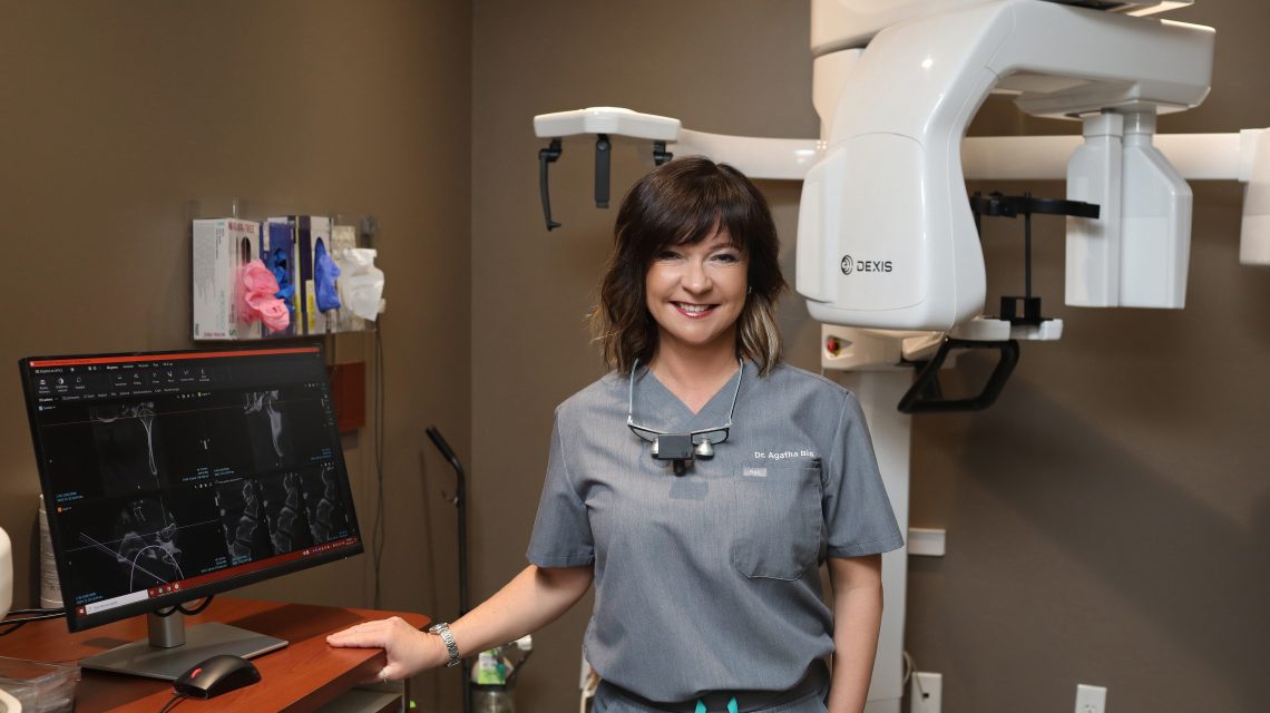
We are excited to announce the integration of cutting-edge technology into our practice with the introduction of a Cone Beam Computed Tomography (CBCT) machine. This state-of-the-art equipment will revolutionize our ability to diagnose and treat TMJ disorders, enhancing precision and efficacy in patient care.
Cone Beam Computed Tomography (CBCT) has become an indispensable tool in the treatment of Temporomandibular Joint (TMJ) disorders due to its ability to provide detailed three-dimensional images of the craniofacial region. By offering high-resolution visualization of TMJ anatomy, including bony structures, soft tissues, and spatial relationships, CBCT enables clinicians to accurately diagnose and assess TMJ pathologies such as erosions, osteophytes, and disc displacement. This precise imaging also aids in treatment planning by guiding the selection of appropriate interventions, monitoring treatment outcomes, and optimizing patient care. In essence, CBCT enhances the precision, efficiency, and effectiveness of TMJ treatment, making it an indispensable tool for clinicians in this field.
Here’s how CBCT contributes to the understanding and management of TMJ disorders:
Visualization of TMJ Anatomy: CBCT provides high-resolution images that allow for detailed visualization of the bony structures, soft tissues, and spatial relationships within the TMJ complex. This includes the condyle, fossa, articular disc, and surrounding structures.
Assessment of Bony Abnormalities: CBCT can identify bony abnormalities such as erosions, osteophytes, fractures, or bony anomalies that may contribute to TMJ pathology.
Evaluation of Joint Morphology: CBCT enables the assessment of TMJ morphology, including the shape, size, and position of the condyle and fossa, which can aid in identifying deviations from normal anatomy.
Analysis of Joint Space: CBCT allows for precise measurement of the joint space, which can be useful in detecting abnormalities such as joint effusion, disc displacement, or degenerative changes.
Assessment of Airway and Soft Tissues: CBCT software evaluates the airway and soft tissues surrounding the TMJ, providing insights into conditions such as airway obstruction or soft tissue pathologies that may be caused by jaw misalignment or contribute to TMJ symptoms.
Treatment Planning: CBCT images aid in treatment planning for TMJ disorders by providing comprehensive information about the underlying pathology, which can guide the selection of appropriate interventions, such as splint therapy, orthodontics, or surgical procedures.
Monitoring Treatment Outcomes: CBCT can be used to monitor treatment outcomes over time by comparing pre- and post-treatment images, allowing clinicians to assess changes in joint morphology and response to therapy.
Overall, CBCT plays a valuable role in the diagnosis, treatment planning, and monitoring of TMJ disorders, providing clinicians with detailed anatomical information essential for delivering effective care to patients.
– Written by Dr. Agatha Bis


